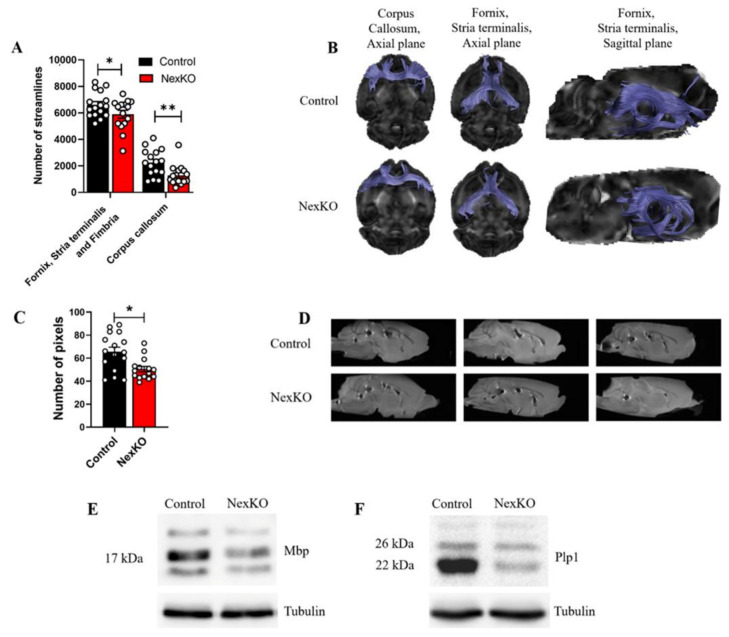
Figure 1.
White matter microstructure, tract connectivity, and myelin deficits in the brains of 1-month-old NexKO mice: (A) Significantly decreased number of streamlines in the limbic outputs through the fimbria/fornix fibers and the stria terminalis as well as the corpus callosum of NexKO mice compared to controls. (B) Brain images overlaid with tractography results for diffusion tensor imaging showing fiber tracking results for control (upper row) and NexKO (lower row) P30 mice. (C) Significantly smaller area of the genu of the corpus callosum of NexKO mice compared to controls. (D) Midsagittal brain images from control and NexKO mice, demonstrating the altered anatomical features of the corpus callosum. Western blots of (E) MBP isoforms and (F) Plp1 expression levels in the cortices of P30 controls and NexKO mice. Data are shown as the mean ± SEM. * p < 0.05, ** p < 0.01; (A,C) two-tailed t-test. (A,C) n = 16 control; n = 15 NexKO.

