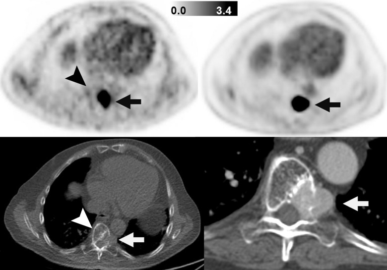
Figure 2:
Images in patient with SDHB-mutant metastatic paraganglioma and pheochromocytoma (patient 1). Images on left are FGln PET (top) and companion CT (bottom) images obtained in single axial thoracic plane. Images on right are FDG PET (top) and companion CT (bottom) images obtained in corresponding axial thoracic plane and were obtained 2 weeks after FGln PET. Hypermetabolic osteolytic spinal metastasis, with extraosseous component, is present (arrows). FGln PET image was obtained 36 minutes after injection of 148 MBq FGln; at this early time point, tracer accumulation in normal bone (eg, unaffected portion of vertebra, arrowheads) is scant.

