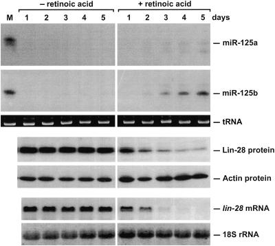
FIG. 8.
Changes in miR-125a and miR-125b levels and lin-28 gene expression following retinoic acid induction of P19 cell differentiation. P19 cells were grown for 5 days in agarose-coated petri dishes in the presence or absence of retinoic acid (1 μM). RNA and protein samples were extracted daily, and equal amounts were analyzed by blotting, using radiolabeled DNA probes to detect specific RNAs (miR-125a, miR-125b, and lin-28 mRNA) and polyclonal anti-Lin-28 antibodies to detect Lin-28. tRNA, 18S rRNA, and actin served as internal standards. Lane M, RNA extracted from 293T cells transiently transfected with either pMIR125a (top pair of panels; detection with an miR-125a-specific probe) or pMIR125b (second pair of panels; detection with an miR-125b-specific probe); note the similar abundance of miR-125b in P19 cells induced for 5 days and in transfected 293T cells. Due to low-level cross-hybridization, miR-125b (faint lower band) was detectable with the miR-125a-specific probe, and miR-125a (faint upper band) was detectable with the miR-125b-specific probe.

